Unveiling Dinosaur Development: The Ontogenetic Growth Stages of Pachycephalosaurus wyomingensis

Introduction to Pachycephalosaurid Ontogeny
In the fascinating world of paleontology, understanding the life cycles of ancient creatures provides profound insights into their biology, behavior, and evolutionary adaptations. One compelling example is the dome-headed dinosaur Pachycephalosaurus wyomingensis, a Late Cretaceous herbivore known for its thick, bony skull dome. This tutorial-style guide explores the growth stages of this dinosaur, drawing from a captivating museum exhibit that illustrates how what were once thought to be three distinct species are actually developmental phases of a single taxon. We’ll delve into the morphological transformations, histological evidence, and taxonomic implications, equipping you with the knowledge to appreciate similar ontogenetic patterns in other dinosaurs.
Ontogeny refers to the developmental history of an organism from its earliest stages to maturity. In dinosaurs, cranial ontogeny—the changes in skull structure over time—is particularly informative due to the preservation of fossilized bones. For Pachycephalosaurus, recent research has revolutionized our understanding by synonymizing two previously named genera, Dracorex hogwartsia and Stygimoloch spinifer, as its juvenile and subadult forms, respectively. This reinterpretation stems from detailed studies at institutions like the Museum of the Rockies, where exhibits showcase these skulls side by side to demonstrate the continuum of growth.
By the end of this post, you’ll learn how to identify key features of each growth stage, interpret histological data, and apply these concepts to broader paleontological questions. Let’s begin with the exhibit that brings this science to life.

Analyzing the Museum Exhibit: A Visual Guide to Skull Growth
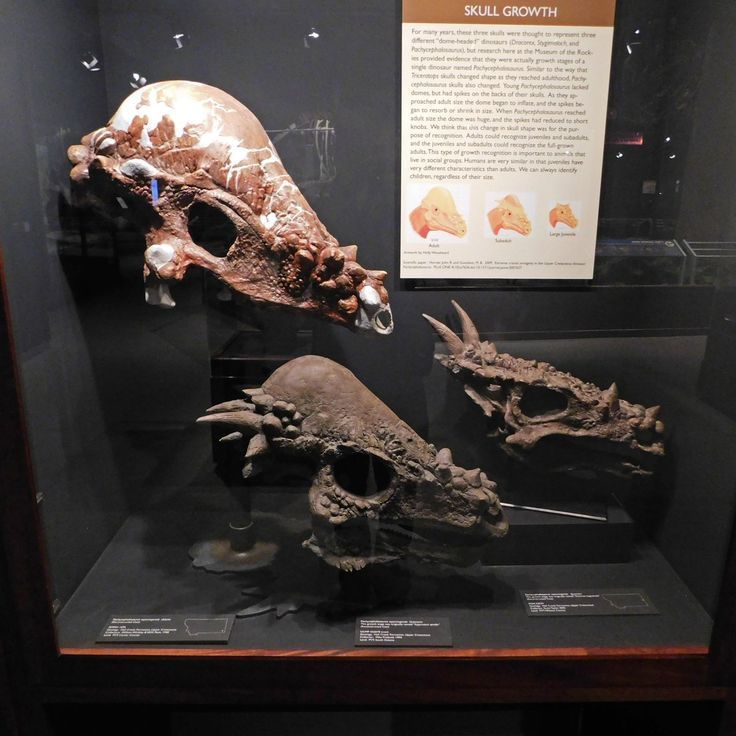
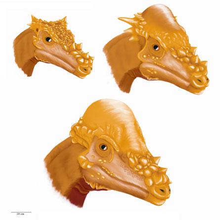
Museum displays, such as the one at the Museum of the Rockies in Bozeman, Montana, serve as educational portals to prehistoric life. The featured exhibit, titled “Skull Growth,” presents three fossilized skulls in a glass case, each representing a different ontogenetic stage of Pachycephalosaurus wyomingensis. Accompanied by an informative placard, the display explains the historical misinterpretation and the current scientific consensus.
Step 1: Identifying the Juvenile Stage (Dracorex hogwartsia)
The smallest skull, positioned on the right in many such displays, is that of a juvenile, formerly classified as Dracorex hogwartsia (meaning “dragon king of Hogwarts,” a nod to its spiky appearance and the Harry Potter series). This specimen, often from South Dakota’s Hell Creek Formation, features a flat, broad skull with prominent spikes and nodes around the squamosals (the bones at the back of the skull) and nasals (near the snout). The frontoparietal dome—the signature thickened area—is minimal or absent, and the skull retains open supratemporal fenestrae (holes behind the eyes). These features give it a “dragon-like” look, but they are transient, designed for rapid growth rather than adult function.
In the exhibit, this skull is labeled with details about its discovery, emphasizing its small size (skull length around 40-50 cm) and the abundance of ornamental spikes, which number in clusters and point backward or outward. To analyze such a fossil in a tutorial context, measure the skull length and note the ratio of dome height to overall cranium: in juveniles, it’s low (less than 1:5), indicating immaturity.
Step 2: Examining the Subadult Stage (Stygimoloch spinifer)
Adjacent to the juvenile is the subadult skull, previously known as Stygimoloch spinifer (“horned demon from the river of death”). Sourced from formations in Montana and Wyoming, this intermediate form shows the beginnings of dome inflation. The frontoparietal region starts to thicken and expand, while the spikes on the squamosals elongate into sharp, curved horns—up to 10-15 cm long—creating a more aggressive silhouette.
Key changes include partial closure of the temporal fenestrae and a lengthening of the skull (typically 60-80 cm). The exhibit highlights how these horns, initially fibrous and vascularized, begin to remodel. In a hands-on tutorial approach, compare the subadult to the juvenile by overlaying diagrams: note the allometric growth where horns grow disproportionately before being resorbed.
Step 3: Observing the Adult Stage (Pachycephalosaurus wyomingensis)
The largest skull, often the centerpiece, represents the mature Pachycephalosaurus wyomingensis. This form boasts a massively inflated dome, up to 25 cm thick, composed of dense bone suited for potential head-butting behaviors. The spikes and horns from earlier stages have eroded into blunt, pyramidal nodes, and the temporal fenestrae are fully closed, resulting in a robust, dome-dominated cranium (skull length exceeding 80 cm).
The placard in the exhibit notes that adults could recognize juveniles through these distinct shapes, aiding social interactions. To tutorialize this, use calipers or digital models to quantify dome volume increase—often tripling from subadult to adult—illustrating how ontogeny facilitates species recognition.
The exhibit’s diagram (showing adult, subadult, and large juvenile silhouettes) reinforces these transitions, with artwork by Holly Woodward depicting the morphological continuum.
Scientific Background: The Hypothesis and Research Timeline
The idea that Dracorex and Stygimoloch are growth stages of Pachycephalosaurus originated from paleontologist Jack Horner’s team at the Museum of the Rockies in the late 2000s. Prior to this, these were described as separate genera based on skull ornamentation: Dracorex in 2006 and Stygimoloch in 1983. However, comparative anatomy and histology revealed shared traits, such as consistent nasal nodes and squamosal landmarks, unifying them into an ontogenetic series.
Step 4: Tracing the Discovery Process
Begin with fossil collection: Specimens from the Hell Creek and Lance Formations (dated 66-68 million years ago) were analyzed. Use stratigraphic data to confirm co-occurrence—Dracorex and Stygimoloch fossils appear in the same layers as Pachycephalosaurus, supporting synonymy rather than temporal separation (though some debate persists).
Next, employ CT scans and thin-sectioning: Coronal scans of subadult domes show open sutures, indicating active growth, while adult domes are fused and dense.
Detailed Morphological Changes: A Step-by-Step Breakdown
Pachycephalosaurids undergo extreme cranial remodeling via metaplasia, a process where fibrous tissue transforms directly into bone.
Step 5: Mapping Cranial Transformations
- Dome Inflation: Starts subtly in juveniles, accelerates in subadults (vascularized tissue adds layers), and peaks in adults (dense, avascular bone for strength).
- Ornament Resorption: Juvenile spikes elongate in subadults, then erode via osteoclastic activity, becoming nodes in adults. Measure horn curvature: 45-60 degrees in subadults vs. blunted in matures.
- Fenestrae Closure: Open in young for flexibility; close progressively, enhancing skull rigidity.
- Skull Elongation: Increases linearly with age, from ~40 cm (juvenile) to >80 cm (adult), following allometric scaling.
Use software like ImageJ for morphometric analysis in your own studies to quantify these ratios.
Histological Evidence: Peering Inside the Bone
Histology provides microscopic proof of growth dynamics.
Step 6: Interpreting Thin Sections
- Juvenile/Subadult: Highly vascularized metaplastic bone with Sharpey’s fibers (indicating keratin sheaths on horns) and erosional surfaces showing remodeling phases.
- Adult: Denser tissue with reduced vascularity, signifying maturity. Compare sections: Subadults have open intrafrontal sutures; adults do not.
Tutorial tip: Prepare slides by embedding fossils in resin, slicing to 30-50 μm, and examining under polarized light to identify fiber orientations.
Implications for Taxonomy and Paleobiology
This ontogenetic model reduces pachycephalosaurid diversity by one-third in some estimates, highlighting over-splitting in dinosaur nomenclature. Behaviorally, the dome’s evolution suggests head-butting in adults for dominance, while juveniles relied on spikes for defense or display.
Step 7: Applying to Other Dinosaurs
Extend this to ceratopsids (e.g., Triceratops frill changes) or hadrosaurids (crest development). Always consider ontogeny before naming new taxa—check for growth series in collections.
Conclusion: Lessons from Ancient Skulls
The Pachycephalosaurus growth exhibit exemplifies how science evolves, turning three “species” into one dynamic life story. By following these steps—from exhibit observation to histological analysis—you can engage deeply with paleontology. Visit the Museum of the Rockies or explore virtual tours to see it firsthand. This understanding not only enriches our view of dinosaurs but also underscores the importance of developmental biology in unraveling evolutionary puzzles.