Pterosaur Fossil Description

The image provides a detailed scientific illustration of fossilized pterosaur bones, likely intended for educational or research purposes. It features multiple views of a pterosaur skull and jaw fragments, accompanied by precise anatomical labels and scale bars, offering a glimpse into the morphology of these ancient flying reptiles. The illustration is monochromatic, with both photographic and line-drawn representations, highlighting the intricate bone structures and their features. This image is a valuable resource for paleontologists, students, or enthusiasts interested in prehistoric life.
Overview of the Illustration
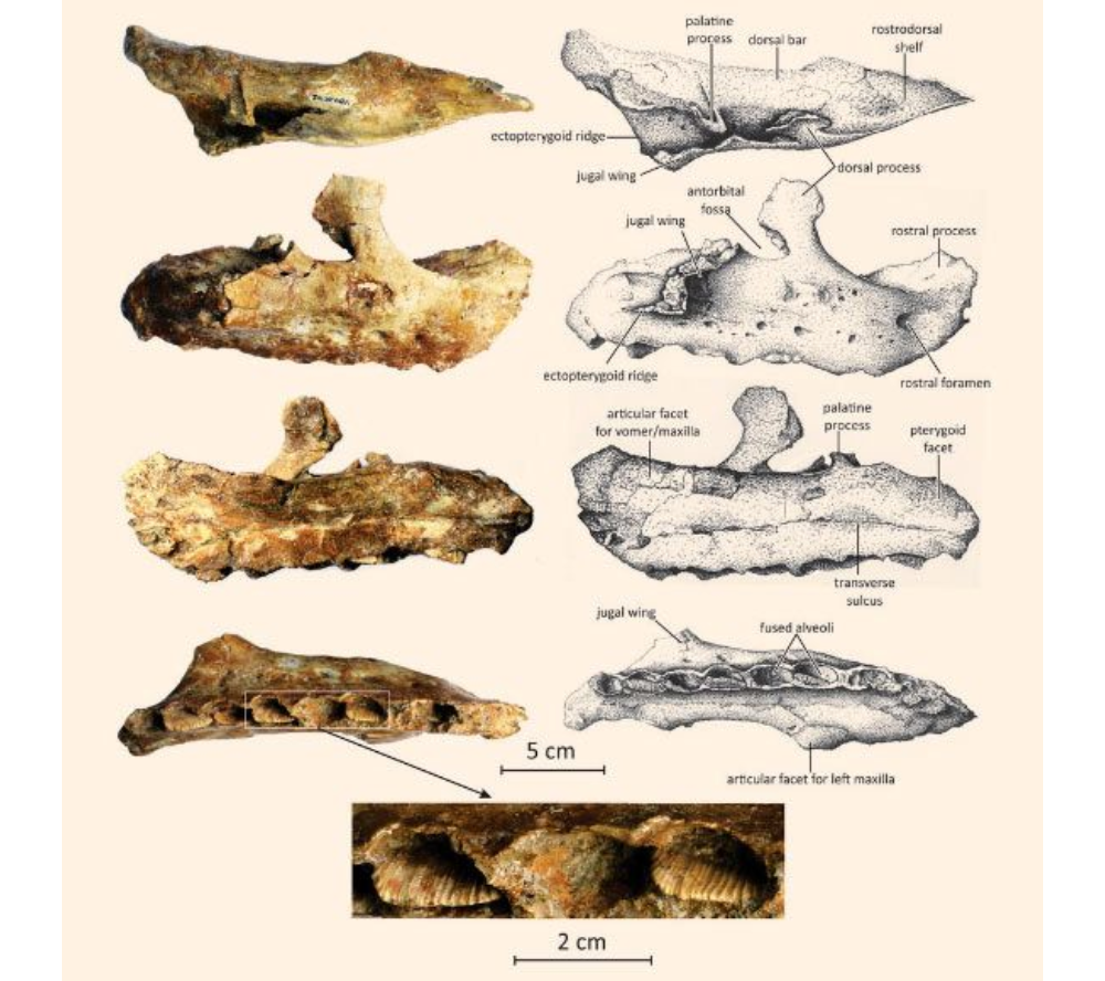
The image is divided into four main sections, each showing different angles of a pterosaur skull and jaw fragments, with a smaller inset at the bottom depicting associated fossilized teeth. Each section includes a line drawing alongside a photograph or shaded rendering, with labels pointing to specific anatomical features. Two scale bars are provided for reference: one indicating 5 cm for the main fossils and another indicating 2 cm for the teeth inset.
Top Section: Lateral View of the Skull
The top section shows a lateral (side) view of a pterosaur skull. The photograph on the left displays the fossil in its preserved state, with visible cracks and a rough texture, indicating its age and the fossilization process. The line drawing on the right labels key features:
- Palatine process: A bony projection involved in the structure of the palate.
- Dorsal bar: A structural element along the top of the skull.
- Rostral process: The forward-projecting part of the snout.
- Ectopterygoid ridge: A ridge associated with the ectopterygoid bone, which connects to the palate.
- Jugular wing: An extension of the jugal bone, contributing to the cheek region.
- Articular facet for maxilla: The surface where the maxilla (upper jaw) articulates with other bones.
Second Section: Ventral View of the Skull
The second section presents a ventral (underside) view of the same skull. The photograph shows the fossil with visible bone sutures and cavities, while the line drawing labels additional features:
- Ectopterygoid ridge: Reappears in this view, emphasizing its role in the palate structure.
- Articular facet for maxilla: Again noted, showing its position from below.
- Palatine process: Visible from the underside, reinforcing its structural importance.
- Rostral foramen: An opening in the snout, likely for nerves or blood vessels.
- Fused premaxillae: The premaxilla bones, which form the tip of the snout, are fused together.
Third Section: Dorsal View of the Jaw
The third section focuses on a dorsal (top) view of a lower jaw fragment. The photograph reveals the fossil’s surface texture, while the line drawing labels:
- Fused alveoli: The sockets where teeth were once anchored, now fused together.
- Transverse sulcus: A groove running across the jaw, possibly related to muscle attachment or nerve pathways.
Fourth Section: Lateral View of the Jaw
The fourth section shows a lateral view of another jaw fragment. The photograph and line drawing highlight:
- Articular facet for left maxilla: The connection point for the left maxilla, indicating the jaw’s role in articulation with the upper skull.
Bottom Inset: Fossilized Teeth
The bottom inset features a small cluster of fossilized teeth, with a 2 cm scale bar for reference. The teeth are elongated and slightly curved, typical of pterosaur dentition, which was adapted for grasping prey such as fish. The photograph shows their preservation state, with visible ridges and a slightly worn surface, suggesting their use during the pterosaur’s life.
Connection to Previous Guides
This pterosaur fossil illustration diverges from the practical tool-focused guides on “12 Essential Woodworking Tools For Beginners,” “Types of Measuring Tools,” and “Welding Tools,” shifting focus to paleontology. However, there’s a subtle connection through the theme of precision and craftsmanship. Just as woodworking, measuring, and welding require accurate tools to shape materials, paleontology relies on precise tools (like calipers and gauges, some of which were featured in the measuring tools guide) to study and reconstruct fossils. For instance, a vernier caliper or measure square could be used to measure the dimensions of these pterosaur bones, ensuring accurate documentation and analysis.
Significance of the Fossil
This illustration likely represents a specimen from a pterosaur species, a group of flying reptiles that lived during the Mesozoic era, alongside dinosaurs. The detailed labeling suggests it’s part of a scientific study, possibly for species identification or understanding pterosaur anatomy. The presence of fused alveoli and specific articular facets indicates adaptations for feeding and flight, key traits of pterosaurs. The scale bars (5 cm for the skull and jaw, 2 cm for the teeth) provide a sense of the specimen’s size, suggesting it may belong to a smaller pterosaur species.
This image, captured as of 06:41 PM EDT on Tuesday, June 03, 2025, serves as an educational tool for those interested in paleontology, offering a window into the ancient world of pterosaurs and the meticulous work involved in studying their remains.