The Elegant Engineering of Viper Fangs and Venom Delivery: Anatomy, Function, and Evolutionary Design in a Post-Fall World

A Comprehensive Tutorial on Viper Venom Systems
Welcome to this in-depth tutorial on the sophisticated anatomy and functionality of viper fangs and venom glands. Drawing from detailed infographics and scientific illustrations, we’ll explore how these structures exemplify precise biological engineering, particularly in the context of adaptation for survival in a challenging environment. This guide is designed for educators, students, biologists, and enthusiasts interested in herpetology, anatomy, or creation science. We’ll break it down step by step, starting with an overview, followed by anatomical details, functional mechanics, and broader implications, all while referencing key biblical insights for a holistic perspective.
Step 1: Understanding the Historical and Conceptual Framework
Before diving into the anatomy, it’s essential to contextualize the design of viper fangs. According to foundational principles in creation science, animals were originally created in a harmonious, vegetarian world as described in Genesis 1:30, where no creature needed predatory mechanisms for survival. However, following the biblical “Fall” (Genesis 3), the world became “cursed,” introducing death, predation, and the need for defensive and offensive adaptations. Vipers, as a prime example, feature specialized fangs and venom systems that appear purposefully engineered to enable survival in this altered ecosystem. These “deadly features” are not random evolutionary accidents but elegantly designed tools for injecting venom into prey, ensuring the snake’s sustenance in a fallen world. This perspective, supported by illustrations from sources like QA International (2009), highlights the intentionality behind such structures, blending biology with theological insight.
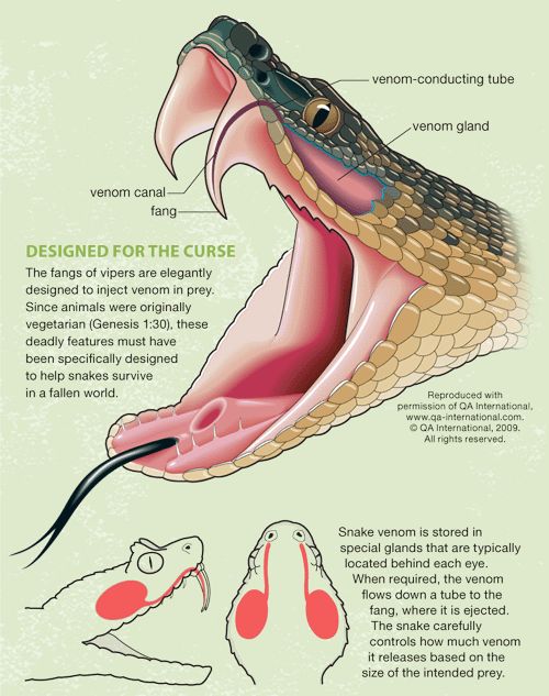
In this tutorial, we’ll treat the viper’s venom system as a model of biomechanical precision, akin to a high-tech injection device. Think of it as nature’s syringe: efficient, controlled, and adaptable. We’ll use the provided infographic as our visual reference, which labels key components like the venom gland, venom-conducting tube, venom canal, and fang.
Step 2: Dissecting the Anatomy – Key Components of the Venom System
Let’s start by examining the core elements illustrated in the infographic. Imagine a viper’s head in mid-strike, mouth agape, revealing the internal plumbing of its venom delivery apparatus. Here’s a detailed breakdown:
- Venom Gland: This is the storage powerhouse of the system, resembling a specialized sac or reservoir. Typically located behind each eye (one on either side of the head for bilateral symmetry), these glands are muscular and compressible. They produce and store venom, a complex cocktail of proteins, enzymes, and toxins tailored to immobilize prey. In vipers, the glands are large relative to the snake’s size, allowing for potent doses. The illustration shows them as purplish-blue ovals, emphasizing their posterior positioning for optimal leverage during a bite.
- Venom-Conducting Tube: Acting as the pipeline, this tube connects the venom gland to the fang. It’s a flexible, durable conduit that ensures venom flows efficiently without leakage. The tube is often embedded in muscular tissue, allowing the snake to contract and propel the fluid forward. In the infographic, it’s depicted as a thin, curving line running from the gland to the base of the fang, highlighting its role in controlled transport.
- Venom Canal: This is the internal channel within the fang itself, a hollow groove or tube that guides venom from the base to the tip. In vipers, fangs are typically grooved or fully canalized (like a hypodermic needle), enabling precise injection. The canal prevents backflow and ensures venom exits only at the point of penetration.
- Fang: The business end of the system, vipers possess long, curved, hinged fangs that fold back when not in use (a proteroglyphous or solenoglyphous dentition). These are replaceable, with new fangs growing in if damaged. The infographic portrays the fang as a sharp, elongated tooth extending from the upper jaw, designed for deep puncture wounds. Unlike non-venomous snakes, viper fangs are evolved for envenomation, piercing flesh and delivering toxin directly into the bloodstream.
To visualize this, refer to the upper illustration: a side-view cutaway of a viper’s open mouth, with labels pointing to each component. The lower diagram simplifies it further, showing paired glands and the flow path to the fangs, underscoring the system’s symmetry and efficiency.
Step 3: Functional Mechanics – How the System Operates Step by Step
Now that we’ve mapped the anatomy, let’s simulate the process as a tutorial walkthrough, as if you’re observing a viper in action. This venom delivery is a marvel of biological control, far more nuanced than a simple bite.
- Venom Production and Storage: The process begins in the venom glands, where specialized cells synthesize venom over days or weeks. Venom composition varies by species (e.g., neurotoxic in some, hemotoxic in others) and is stored under pressure in the glands. The snake can regenerate venom after depletion, but this takes time—typically 1-3 weeks for full replenishment.
- Triggering the Strike: When hunting or defending, the viper strikes with lightning speed (up to 2-3 meters per second). As the mouth opens, the fangs rotate forward from their folded position, powered by jaw muscles.
- Venom Flow Activation: Muscles surrounding the venom glands contract, forcing venom through the conducting tube. This is not an all-or-nothing release; the snake modulates pressure based on prey size and threat level. For small prey, it might use minimal venom to conserve resources; for larger threats, a full dose ensures quick immobilization.
- Injection via the Fang: Venom enters the canal and exits through the fang’s tip upon penetration. The infographic notes: “When required, the venom flows down a tube to the fang, where it is ejected.” Depth of bite and angle are critical—vipers aim for vital areas to maximize effect. The snake “carefully controls how much venom it releases based on the intended prey,” demonstrating cognitive regulation.
- Post-Bite Recovery: After striking, fangs retract, and the snake tracks its envenomated prey, which succumbs to paralysis, tissue damage, or cardiac arrest. This system minimizes energy expenditure, allowing the viper to survive in sparse environments.
Pro Tip: In a lab or educational setting, models or 3D animations can replicate this. For safety, always study via diagrams—never handle live vipers without expert supervision.
Step 4: Broader Implications and Adaptations
From a tutorial standpoint, the viper’s venom system teaches us about biomechanical efficiency and adaptation. In a “fallen world” framework, these features represent post-creation modifications for survival amid predation and scarcity. Scientifically, vipers’ designs inspire medical advancements, like antivenoms or painkillers derived from toxins.
Comparisons:
- Vs. Other Snakes: Cobras have fixed fangs and spray venom, while vipers emphasize injection for close-quarters hunting.
- Evolutionary vs. Design Perspective: While evolutionary biology posits gradual development from non-venomous ancestors, the creationist view sees this as specific, curse-adapted engineering.
Safety Note: If encountering vipers, recognize symptoms of envenomation (swelling, nausea) and seek immediate medical help.
Conclusion and Further Resources
This tutorial has unpacked the viper’s fangs and venom system as a testament to intricate design, from glandular storage to precise ejection. For deeper study, consult sources like QA International’s illustrations (reproduced with permission, © 2009) or biblical texts like Genesis. Explore herpetology texts or online simulations for interactive learning. If you’re creating content for a website, embed similar infographics to enhance engagement—remember, visuals like this one make complex biology accessible and fascinating.
Feel free to comment below with questions or share your own observations on serpentine adaptations!